
Detailed explanation of the microscope knowledge points of the junior high school biological high school entrance examination review
The imaging principle of the microscope:
when the light shines on the mirror, it will pass through the shutter and the through hole, and then be projected on the transparent specimen. The objective lens magnifies the specimen for the first time to form an inverted real image. This real image passes through the lens barrel and is enlarged by the eyepiece for the second time to form an inverted virtual image, which is finally presented in our eyes.
2, the relationship between lens length and magnification:
(1) The length of the eyepiece is inversely proportional to the magnification, that is, the longer the eyepiece, the smaller the magnification.
(2) The length of the objective lens is proportional to the magnification. The longer the objective lens, the larger the magnification.
3, in the high magnification objective lens, the observed field of view is small and the light is darker
but the cells appear large and clear, and the number is relatively small; while in the low magnification objective lens, the field of view is broad and bright, and the cells appear small and blurry, and the number is large.
4, with the increase of the magnification of the objective lens, the distance between the objective lens and the slide will gradually decrease
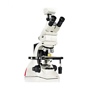
In addition, the structure of the microscope also includes the objective lens, lens barrel, converter, stage, through hole, tablet clip and shutter and other components. Among them, the objective lens is a lens close to the slide, responsible for magnifying the object; the lens barrel is used to connect the eyepiece and the converter; the converter is equipped with different multiples of the objective lens, so as to exchange according to need; the stage is used to place the slide specimen; The light hole is located in the center of the stage to ensure that light can pass through; the tableting clip is used to fix the slide specimen; and the aperture on the shutter can adjust the light intensity to adapt to different observation needs.
 Microscope skills in detail: from basic to advanced applicat
 A complete analysis of the imaging principles and skills of
 Detailed Explanation of Microscope Imaging Characteristics a
 Here are 20 trivias you didn't know about microscopes!