
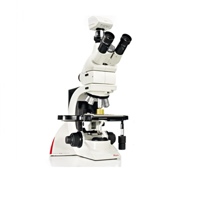
Stereo microscope Stereo microscope Dissecting microscope
Comparison of stereomicroscopes with ordinary biological microscopes
Features and usage of stereomicroscopes
Objects under stereomicroscopes are not inverted. Usually, light shines on the specimen obliquely from the object above, so the surface of the specimen is observed. Compared with ordinary biological microscopes, stereomicroscopes have a larger depth of focus and can place larger samples, such as stems, leaves, flowers and other plant organs. Observers can also perform dissection operations under stereoscopes. Its magnification is the product of objective lenses, variable-magnification objectives and eyepieces. However, objective lenses are generally not used. Once the objective lens is used, the field of view observed is small, the depth of focus is also small, and the light in the field of view is also dimmed.
• Characteristics of stereomicroscopes
1. The left and right beams of light in the binocular tube are not parallel, but have a certain angle - the volume viewing angle (generally 12 degrees - 15 degrees), so the imaging has a three-dimensional sense;
2. Like upright, easy to operate and dissect, which is due to the lens under the eyepiece to reverse the image;
3. The working distance is long and the field of view diameter is large.
4. The depth of focus is large, which helps to observe the whole layer of the object to be inspected.
• How to use the stereo microscope
1, According to the color of the object, select the black and white side of the workbench, and place the object to be observed on the slide or petri dish and then place it on the workbench.
2, choose the appropriate magnification, replace the required eyepiece (10 or 20). If observed below 80, 2 large objective lenses can be removed, and the effective working distance is 87mm. If 2 large objective lenses are added, the magnification can reach 160, and the effective working distance is 26mm. Adjust the working distance and release the locking handwheel. It is achieved by pulling out or pressing into the movable pillar.
3, when operating, move the object to the center position of the table plate, rotate the lifting handwheel, so that the left eyepiece can see a clear object image. If the image of the right eyepiece is not clear, the eyepiece focusing ring can be rotated to obtain a clear object image as the left eyepiece, so that a clear object image with a three-dimensional sense can be seen, and the focusing work is basically completed. In order to obtain an appropriate magnification, it can be achieved by turning the magnification adjustment ring to change the magnification of the variable magnification objective. The magnification of the zoom objective can be read on the reading circle. If necessary, the right-angle prism group can be adjusted to change the distance between the eyepieces to fit the pupil distance above the observer's eyes. Loosen the tight screw to rotate the microscope around the axis at any position.
 Microscope skills in detail: from basic to advanced applicat
 A complete analysis of the imaging principles and skills of
 Detailed Explanation of Microscope Imaging Characteristics a
 Here are 20 trivias you didn't know about microscopes!